Banghart Lab
  • Home
  • Research
  • Publications
  • People
  • Resources
  • Contact
  • Links
  • Open positions
  • Home
  • Research
  • Publications
  • People
  • Resources
  • Contact
  • Links
  • Open positions
Random, hopefully Useful stuff that we have developed in the lab. Some published, some not... 
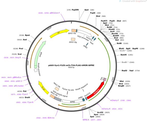
Photoactivatable or "caged" neuropeptides and small molecules

We have developed a suite of light-sensitive pharmacological probes for stimulating and inhibiting endogenous neuropeptide receptors with light. This work started during Matt's postdoc in Bernardo Sabatini's lab and is being continued at UCSD. Although initial progress was made on opioid receptors, the lab is interested in other neuropeptides as well. Sample requests, or interest in collaborating to develop additional caged molecules, should be directed to Matt by email. NIDA funded researchers can obtain milligram quantities of several reagents through the NIDA Drug Supply Program (NDSP) for free. We are also happy to consult on appropriate optical configurations for uncaging and experimental design to avoid common pitfalls associated with using these reagents. ​
opioids
enkephalin
dynorphin
DAMGO
oxymorphone
naloxone
other neuropeptides
substance P
gastrin-releasing peptide
oxytocin
cholecystokinin

All of the reagents developed thus far are activated by ultraviolet or violet light (350-405 nm) but not longer wavelengths used for imaging green and red fluorophores. This is both an advantage (imaging-compatible) and a disadvantage (specialized light sources required).

More detailed information is available below.
Opioid receptor ligands
We chose to photosensitize the endogenously occurring opioid neuropeptide leucine-enkephalin  due to its abundance in the brain, nanomolar efficacy for activating both mu and delta opioid receptors, and because it is more chemically stable than the more abundant but pharmacologically similar analog methionine-enkephalin. We also photosensitized the kappa agonist dynorphin 8, which is the shortest and most abundant form in the brain (it also activates mu and delta with similar affinity to kappa). We initially developed variants of leucine-enkephalin and dynorphin 8 that can photoreleased with 355 nm UV light using classic nitrobenzyl caging groups placed on the N-terminal tyrosine side chain (CYLE & CYD8, Banghart & Sabatini, Neuron, 2012). We then further improved inactivity at the high-affinity delta receptor in the caged form by moving the caging group to the N-terminal amine, and switched to dimethoxynitrobenzyl-derived caging groups to shift the wavelength sensitivity out to 405 nm (N-MNVOC-LE, Banghart, He & Sabatini, ACS Chemical Neuroscience, 2018). This allows both mu and delta receptors to be probed simultaneously. Importantly, these reagents are not photoreleased by longer wavelength blue light that is used for imaging GFP, but respond well to commercial UV LEDs. Although not included in that paper, we extended this optimization to dynorphin 8 as well (N-MNVOC-Dyn8). Notably, because the affinity reduction at delta resulted from masking the charged amino terminus in a neutral carbamate linkage to the caging group, photorelease involves a decarboxylation step that slows the rate of photorelease to the timescale of hundreds of milliseconds, which is just a bit slower than GPCR-effector activation (e.g. GIRKs). If the application is to measure receptor or effector activation kinetics, the less inactive but faster-releasing tyrosine-caged variants may be most optimal. 

​Although the enkephalin and dynorphin variants developed to date have not been validated for 
in vivo applications, we developed CNV-Y-DAMGO, PhOX (caged oxymorphone), and PhNX (caged naloxone) with in vivo applications specifically in mind. CNV-Y-DAMGO, which is a peptide and thus poorly penetrates the blood-brain barrier, can be administered through an (optofluidic) cannula and photoactivated via an implanted optical fiber in vivo. PhOX and PhNX are relatively hydrophobic small molecules and can be administered systemically (e.g. subcutaneous injection) and photoactivated in vivo through an optical fiber. Manuscripts describing CNV-Y-DAMGO and PhOX/PhNX are forthcoming. ​

The following table summarizes the key properties of each variant we have developed. Potencies were determined at human receptors expressed in HEK293 cells using a functional SEAP assay. In brain slices, the peptides are ~10x less potent, likely due to peptidase activity in tissue. We typically recirculate 1-20 µM, depending on the application. Please contact Matt for further information.
Name
Caged variant of:
Wavelength sensitivity
Mu EC50
​(LE=90 nM, D8=63 nM, DAMGO=1.5nM)
Delta EC50
​(LE=3 nM, D8=10 nM)
Kappa EC50
​(LE inactive, D8=7 nM)
Photolysis kinetics (approx)
Available through NDSP?
Reference
CYLE
leu-enkephalin
355 nm
16 µM
1.7 µM
inactive
10's of µs
yes
​Neuron 2012
CYD8
dynorphin 8
355 nm
23 µM
3.9 µM
16 µM
10's of µs
yes
Neuron 2012
CNV-Y-LE
leu-enkephalin
355-405 nm
12 µM
0.4 µM
inactive
10's of µs
yes
​ACS Chem Neuro 2018
N-MNVOC-LE
leu-enkephalin
355-405 nm
34 µM
17 µM
inactive
hundreds of ms
no
ACS Chem Neuro 2018
N-MNVOC-D8
​dynorphin 8
​355-405 nm
41 µM
28 µM
33 µM
hundreds of ms
no
unpublished
CNV-Y-DAMGO
DAMGO
355-405 nm
1.7 µM
N.D.
N.D.
​10's of µs
no
bioRxiv
PhOX
oxymorphone
​355-405 nm
>100 µM 
>100 µM
>100 µM
​10's of µs
no
​bioRxiv
PhNX
naloxone
355-405 nm
​>100 µM
​>100 µM
​>100 µM
10's of µs
no
bioRxiv
These molecules have been validated using either a functional SEAP or GloSensor cAMP assay in HEK293 cells transfected with opioid receptors, slice electrophysiology at endogenous receptors (Mu receptor mediated GIRK current activation, and mu and delta receptor-mediated suppression of synaptic transmission), and more recently, genetically-encoded optical neuropeptide sensors. 

​It is important for potential users to realize that "caged" molecules are not necessarily biologically dead prior to photolysis, and that caging peptides is generally more difficult than caging a small molecule drug or neurotransmitter, as peptides are large and contain several sites that interact with receptors. Thus caging a single site, which affords clean photo-release, typically only attenuates activity rather than abolishing it completely. In general, an affinity attenuation of 500-fold is sufficient to yield complete receptor activation at a fully inactive concentration of caged peptide though this depends on the steepness of the dose-response curve. This residual activity limits the concentrations that can be applied to tissue to tens of micromolar concentrations. The solubility of hydrophobic peptides at tens of micromolar and higher concentrations in saline solution can also be limiting. Thus there is often a narrow concentration window in which caged peptides can be photoconverted from a completely inert state to one that saturates receptors. 
About the antagonists
We have developed two variants of the broad-spectrum opioid receptor antagonist naloxone: CNV-NLX and PhNX (photoactivatable naloxone). CNV-NLX works great in brain slices, but does not penetrate the blood-brain barrier, as it is constitutively charged. PhNX, on the other hand, can be administered systemically (e.g. subcutaneous injection) and photoactivated in vivo through an optical fiber, like the caged agonist PhOX. Both molecules ncorporates a dimethoxycarboxynitrobenzyl-derived caging group and thus responds to 405 nm light. 

To characterize CNV-NLX, we collaborated John Williams at OHSU, (Banghart et al, Molecular Neuropharmacology, 2013, see also Williams JT, Molecular Neuropharmacology, 2014). 

A manuscript describing PhNX (along with PhOX) is forthcoming. The key difference between CNV-NLX and PhNX is that PhNX works well in vivo whereas CNV-NLX doesn't get into the brain very well. 
Other peptides
We have also developed caged variants of Substance P, CCK(8S), GRP(14-27), and Oxytocin. A manuscript describing a novel caging strategy and validated reagents is currently in preparation. The caging strategy is highly general and can be used to produce caged variants of many neuropeptides and peptide antagonists. Please contact Matt for more information. 

Viruses and plasmids

pAAV-Syn1-FLEX-mCh-T2A-hMOR-WPRE (Addgene 166970)
​

Cre-dependent human mu opioid receptor (Oprm1) with stoichiometric expression of volume-filling mCherry


This reagent has been used to overexpress MOR in genetically-targeted neurons and to rescue MOR expression in knock-out mice.
​
He XJ, Patel J, Weiss CE, Ma X, Bloodgood BL, Banghart MR. Convergent, functionally independent signaling by mu and delta opioid receptors in hippocampal parvalbumin interneurons. https://doi.org/10.1101/2021.04.23.441199 

Liu, S., Kim, D., Oh, T.G., Pao, G.M.M., Kim, J., Palmiter, R.D., Banghart, M.R., Lee, K.F., Evans, R.M. and Han, S., “Neural basis of opioid-induced respiratory depression and its rescue,” Proceedings of the National Academy of Sciences (2021), June 8, 118(23):e2022134118. doi: 10.1073/pnas.2022134118.

Picture

Resolving GFP from YFP (both optically and genetically)

A filter cube for imaging GFP without contamination from YFP:
     excitation: 452/25
     dichroic: 484 LP
     emission: 494/20
All filters are available from Semrock for 25 mm cubes.
This cube works well with various GFP strains in Cre/Ai32 (ChR2-eYFP) crosses for targeted patching of GFP cells and optogenetic stimulation of Cre-expressing axons (see Figure 7 in Banghart & Neufeld et al Neuron 2015). A decent camera may be required as the GFP signal is a bit dim relative to a standard eGFP cube. Although the excitation filter gets a bit of YFP, by omitting the peak of GFP's emission spectrum, the emission filter almost completely rejects any YFP signal.
Picture


On a related note, generic primers against GFP may give strong bands against YFP when the Ai32 transgene is present due to their high sequence similarity. We generated primers for GFP that do not recognize YFP:
     forward: CTG ACC TAC GGC GTG CAG TGC TT
     reverse: GCT CAG GGC GGA CTG GGT G
     product band ~ 500 bp
​Thermocycler protocol:​   
     Step     Temp     Time    Note
       1            94          3m
      2            94          45s
      3            66          45s       1.0*/cycle
      4            72           45s
      5                                       repeat steps 2-4 for 8 cycles
      6            94          30s
      7            58           30s
      8            72           30s
      9                                      repeat steps 6-8 for 19 cycles
     10           72           10m
     11           10                        hold
Proudly powered by Weebly